With the three-dimensional cell atlas to precision medicine
The US National Institutes of Health NIH is funding the creation of a three-dimensional map of the human lymphatic system. Bernd Bodenmiller, Professor for Quantitative Biomedicine at the University of Zurich and ETH Zurich and a pioneer in imaging mass cytometry, plays an important role in this: a visit to the Bodenmiller Lab.
The building of the Institute for Quantitative Biomedicine is the newest at the University of Zurich Irchel. It was only occupied last September. It stands at the very top of the campus, offers a lot of light and a view of the Zürichberg forest. A bright red, curved staircase leads up to the top floor. The simple offices of the professors, which are lined up here along a wide corridor, resemble aquariums somewhat, however, with two completely glazed fronts facing the corridor and the outside. The glass wall and door of Bernd Bodenmiller’s office, though, is covered with large-format, colourful pixel paintings: beautiful images of cells produced using what is known as imaging mass cytometry, or IMC.
Bodenmiller is a specialist in this process. He developed it in 2014 together with Detlef Günther, Professor for Trace Element and Micro Analysis at ETH Zurich. Today, the Bodenmiller Lab is known as the world leader in IMC and 3D analysis – and is playing a key role in a current research project of the US National Institutes of Health NIH: the creation of a 3D map of the organs of the lymphatic system with the help of this expertise. The project is coordinated by Mark Atkinson of the College of Medicine at the University of Florida. It received funding for four years and is to develop three-dimensional tissue maps for the three most important lymphoid organs of the human body: the spleen, the thymus and the lymph nodes.
Connected across the globe
The project is part of a larger endeavour: the NIH envisions to measure the entire body of a healthy human being on the single cell-level with the novel methods. It is called Human BioMolecular Atlas Program, in short HuBMAP. Participants are 18 different, leading research teams from the US and Europe who, in turn, collaborate with many researchers from around the world. The images are all to be stored in the same format in one database and made available to researchers across the globe.
The lymphatic system plays a central role, as it has key physiological functions within the healthy body: it ensures the removal of excess body fluids, absorbs and transports fatty acid into the bloodstream and it filters the blood. It produces and activates immune cells and hence builds up the primary defence against infections and cancer. And it creates and activates so-called regulatory immune cells that serve as protection against autoimmune diseases.
«It is like the difference
between a blurry black-and-white image
and a high-resolution colour image.»
This is not the first collaboration between Bernd Bodenmiller and Mark Atkinson. Their cooperation goes way back to the beginnings of the IMC method – and its echo in the professional community. The young German describes it as follows: «We had just developed the IMC imaging method, had it published and started to undertake various further developments of the methodology when I received a call from an NIH Grant Coordinator. He asked me whether I was interested in co-applying for an NIH Grant.» One must know that, not like it is customary in Europe, it is the NIH Grant Coordinators’ task to actively suggest researchers that could make good group members within a project. They practice active team building in order to set up ideal groups to reach the requirements or main objective of a project.
Suitable partners
This first project was on diabetes. Within the scope of an NIH project single-cell analyses of islet cells in type 1 diabetes should be made using high-multiplex imaging. Mark Atkinson is a clinician and has established the world’s largest human biobank for pancreatic tissue from type 1 diabetes donors in the USA. Bodenmiller mainly researches tumours. He analyses cancer tissue with his imaging procedure. The insights thereof are to enhance treatment possibilities. Bodenmiller recounts: «The Coordinator said, ‘Mark has the samples, you have the technology, and you can further develop this within the project.’» Together with a group from Geneva Mark was already familiar with, they successfully applied for the project. This is how the cooperation came about. «It went very well,» Bodenmiller emphasises.
By now, the company Fluidigm has put the IMC technology as a product on the market. And Bodenmiller again collaborates with Atkinson to map the lymphatic system. «Thanks to his biobank Mark has a large number of samples from the human lymphatic system – and at the same time we have a great interest in the immune and lymphatic system in connection with out cancer research,» the biochemist explains. This is why the two of them handed in a joint application for the second time and were successful again.
Blue light without siren
The devices used for the research pale in visual comparison to their global reputation – the most eye-catching aspect are their partly bright orange casings. They consist of a mass analysis device, the CyTOF, and a box inside which the laser ablation system is running.
Three of these machines are lined up behind each other in the group’s lab. During our visit, one of the machines is measuring immune cells. It hardly makes any noise. Only a blue lamp that briefly and silently flashes like the blue light of a police car gives evidence to something happening. Promptly, Sujana Sivapatham, one of the scientific members of staff, appears in the lab and immediately sits down at the computer next to the machine. The young woman explains that the flashing signal gets transmitted to the computer on her desk.
«I only have one brain –
but luckily, I have 29 group members
who are at least just as smart as I am.»
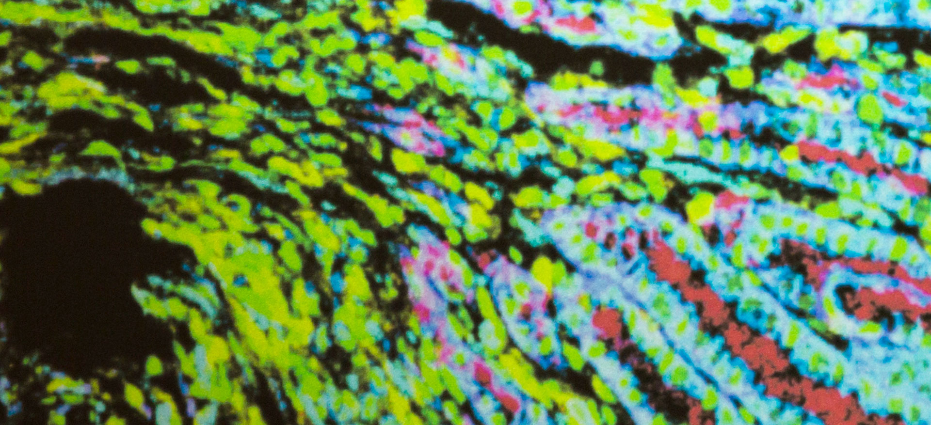
So, what exactly is taking place inside the machines? The scientist offers a simple explanation: they work with tissue samples that are only a few micrometres thick. So-called antibodies are applied to the tissue, with the help of which certain markers can be detected in the tissue. The antibodies were labelled with different metal isotopes beforehand. The researcher then describes the process like this: «We can measure metals in the machine. The laser shoots at the tissue point by point and removes and measures it. In the end, what remains are pixels like on a screen with information of more than 40 markers which we then put together to form a picture.»
The advantage of this system compared to traditional measurement techniques is that researchers can currently look at up to 50 markers, possibly up to 100 markers in the future (and not only three to five like with the standard methods). This provides a whole other depth of information. For example, you not only see that there is an immune cell but also what it is doing right now. «It is like the difference between a blurry black-and-white image and a high-resolution colour image,» Bodenmiller says.
A walk-in tumour
Using this method, three-dimensional models can also be created by making multiple cuts in the tissue which are then measured layer by layer and assembled at the computer to form a 3D model. Bodenmiller’s group is part of a consortium that develops a virtual reality in which 3D models can be researched. For example, a cancer tumour can be depicted in such a way that one can walk into it and look at and analyse its interior.
The project on the three-dimensional map of the lymphatic system ends in June. Have they mapped everything? The professor laughs: «No, we have not yet measured it all. But we are currently applying for an extension of the project. One of the main objectives was to create the fundamentals for large-scale measurements.» In fact, much data was generated for instance from the lymph node. «However, we also have to accept the fact that while we are able to measure a great amount with today’s technologies the organs are still huge compared to the measurement parameters, as we are measuring in the dimension of cubic millimetres,» Bodenmiller states. This is why the researchers put great effort into defining the most interesting regions and measuring those. But much remains to be done.
A group of top people
At the same time, the group of Bernd Bodenmiller participates in no less than four additional international research projects. The Horizon 2020 programme IMMUcan works on defining groups of different cancer cells and immune environments of cancer patients by means of images in order to enable personalised treatments. The MaCaROM Project within the framework of a Marie Skłodowska-Curie Grant is dedicated to the analysis of how diverse breast cancer cells in breast cancer organoids react to different drugs. In a second Marie Curie project called Spatial Organoids three-dimensional organoid cultures are assessed over time to find out which molecular and spatial factors define the heterogeneity of breast cancer cells. This, too, shall help to improve the targeted treatment of breast cancer. The fourth project is funded by the ERC and called Precision Motifs; it analyses breast cancer metastases.
Isn’t he at risk of merely acting as the administrator of this many tasks at the same time? The Group Leader denies: «The art is to have great co-workers who carry and implement the projects with a lot of personal responsibility and enthusiasm. »Indeed, currently four of the 29 staff members were awarded with a scholarship. Bodenmiller’s explanation: the people who apply for a position within his group have the ambition to make a difference and at the same time create a good basis for their next career step. This is why they write research proposals for Marie Curie Grants and other funding opportunities. And Bodenmiller adds: «I am very happy that my staff members are so successful in this.» His brain would have to be thirty times its size if he were to steer every project in the smallest of details, he says. «I only have one brain – but luckily, I have 29 group members who are at least just as smart as I am.»
The biochemist finds it hard to decide on one favourite project: «I have the luxury of realising the projects that interest me. Of course, sometimes there are projects with a lot of bureaucracy. But as soon as a project gets to the stage where results can be generated, it’s great fun.»
However, he knows exactly what he is most proud of: that he and his group successfully developed a technology that is now used in many projects and by a large number of other people. That this technology has a real impact and is used in a clinical context. «It’s brilliant that things we have developed are getting to the patient,» he states.
Interview with Bernd Bodenmiller (in german)

Bernd Bodenmiller
Bernd Bodenmiller has been Associate Professor for Quantitative Biomedicine at the University of Zurich and ETH Zurich since 2020. Originally from Germany, he studied Biochemistry at the University of Bayreuth and at ETH Zurich. He then worked as a doctoral student in the Systems Biology Lab of Prof. Ruedi Aebersold at ETH Zurich and received his PhD in Systems Biology in 2008. The following year, Bodenmiller accepted a postdoctoral position in the lab of Prof. Garry P. Nolan at Stanford University, USA, where he worked until 2012. In 2013, he was appointed SNSF Assistant Professor at the Institute of Molecular Biology at the University of Zurich. Since 2019, he has been the Founding Director of the Institute of Quantitative Biomedicine at the University of Zurich. Bernd Bodenmiller is married, has two children and lives in Zurich.


NIH-Projekt
HuBMAP: A 3D Tissue Map of the Human Lymphatic System
- Programme: NIH Subaward
- Duration:14. September 2018 – 30. June 2022
(45.5 months) - Contribution for University of Zurich: 214’898 $